 Lower thalamic susceptibility, higher susceptibility of the globus pallidus linked to clinical disability
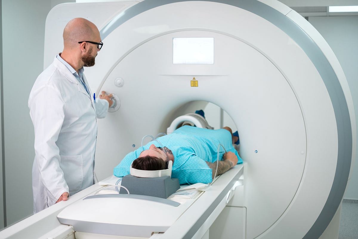
MONDAY, Sept. 24, 2018 (HealthDay News) — Brain iron at quantitative magnetic resonance imaging (MRI) is associated with disability in multiple sclerosis (MS), according to a study recently published in Radiology.
Robert Zivadinov, M.D., Ph.D., from the University at Buffalo in New York, and colleagues conducted a prospective study between March 2009 and November 2013 involving 600 participants with MS (452 with relapsing-remitting MS and 148 with secondary progressive MS) and 250 age- and sex-matched healthy controls. Magnetic susceptibility was measured using 3.0-T MRI.
The researchers found that participants with MS presented with lower thalamic susceptibility (−7.5 versus −1.1 ppb) and higher susceptibility of basal ganglia (62 versus 54.8 ppb), compared with control participants. There was a correlation for lower thalamic susceptibility with longer disease duration, higher degree of disability, and secondary progressive course (β = −0.42, −0.64, and −4.3, respectively). There was a correlation for higher susceptibility of the globus pallidus with higher disability (β = 2). Lower thalamic susceptibility and higher susceptibility of the globus pallidus remained associated with clinical disability after correction for each individual structural volume in voxelwise analysis.
“In this large cohort of MS patients and healthy controls, we have reported, for the first time, iron increasing in the basal ganglia but decreasing in thalamic structures,” Zivadinov said in a statement. “Iron depletion or increase in several structures of the brain is an independent predictor of disability related to MS.”
Several authors disclosed financial ties to the pharmaceutical and medical device industries.
 Pre-Prostate Biopsy MRI Use Increased From 2017 to 2026
  PET/MRI Offers Diagnostic, Prognostic Value in Pediatric Epilepsy
  Dense Breast Notification Linked to Increase in Use of Supplemental Ultrasound
  Nighttime Light Exposure Linked to Adverse Cardiac Remodeling
  Cardiac Remodeling May Portend Cancer Risk
  Volume Rendering of Myocardium, Cardiac Valves Enables Visualization
  CMR-Modeled PCWP Independent Risk Factor for Heart Failure
  Non-MRI-Conditional ICDs Still Function After MRI