


 MRI can be performed safely in patients with non-MRI-conditional devices, including pacemakers
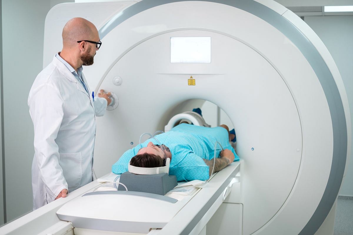
FRIDAY, Oct. 23, 2020 (HealthDay News) — Magnetic resonance imaging (MRI) can be safely performed in patients with non-MRI-conditional cardiac devices, including pacemakers and abandoned leads, according to a study published online Oct. 22 in Radiology: Cardiothoracic Imaging.
Sanjaya K. Gupta, M.D., from Saint Luke’s Mid-America Heart Institute in Kansas City, Missouri, and colleagues examined the safety and clinical utility of MRI in participants with non-MRI-conditional cardiac implantable electronic devices. A total of 532 patients were enrolled in the Patient Registry of Magnetic Resonance Imaging in Non-Approved Devices registry and had device interrogations performed before and after each MRI.
Cardiac devices included pacemakers, implantable cardioverter defibrillators, cardiac resynchronization therapy (CRT) pacemakers, CRT defibrillators, and abandoned leads (46, 30, 4, 17, and 2 percent, respectively). The researchers found that of all MRI examinations, pacemaker-dependent patients comprised 27 percent. There were no reports of patient- or device-related complications. One hundred fifty physicians completed clinical utility surveys of MRI examinations. These MRI examinations changed suspected diagnosis 25 percent of the time, changed suspected prognosis in 26 percent of patients, and changed planned medical or surgical treatment 42 percent of the time.
“This study also offers support for the extensive nonphysician resources necessary to perform these MRI examinations safely by identifying a substantial impact on clinical care in a large proportion of surveyed cases,” the authors write.
 Pre-Prostate Biopsy MRI Use Increased From 2017 to 2026
  PET/MRI Offers Diagnostic, Prognostic Value in Pediatric Epilepsy
  Dense Breast Notification Linked to Increase in Use of Supplemental Ultrasound
  Nighttime Light Exposure Linked to Adverse Cardiac Remodeling
  Cardiac Remodeling May Portend Cancer Risk
  Volume Rendering of Myocardium, Cardiac Valves Enables Visualization
  CMR-Modeled PCWP Independent Risk Factor for Heart Failure
  Non-MRI-Conditional ICDs Still Function After MRI


.jpg)