


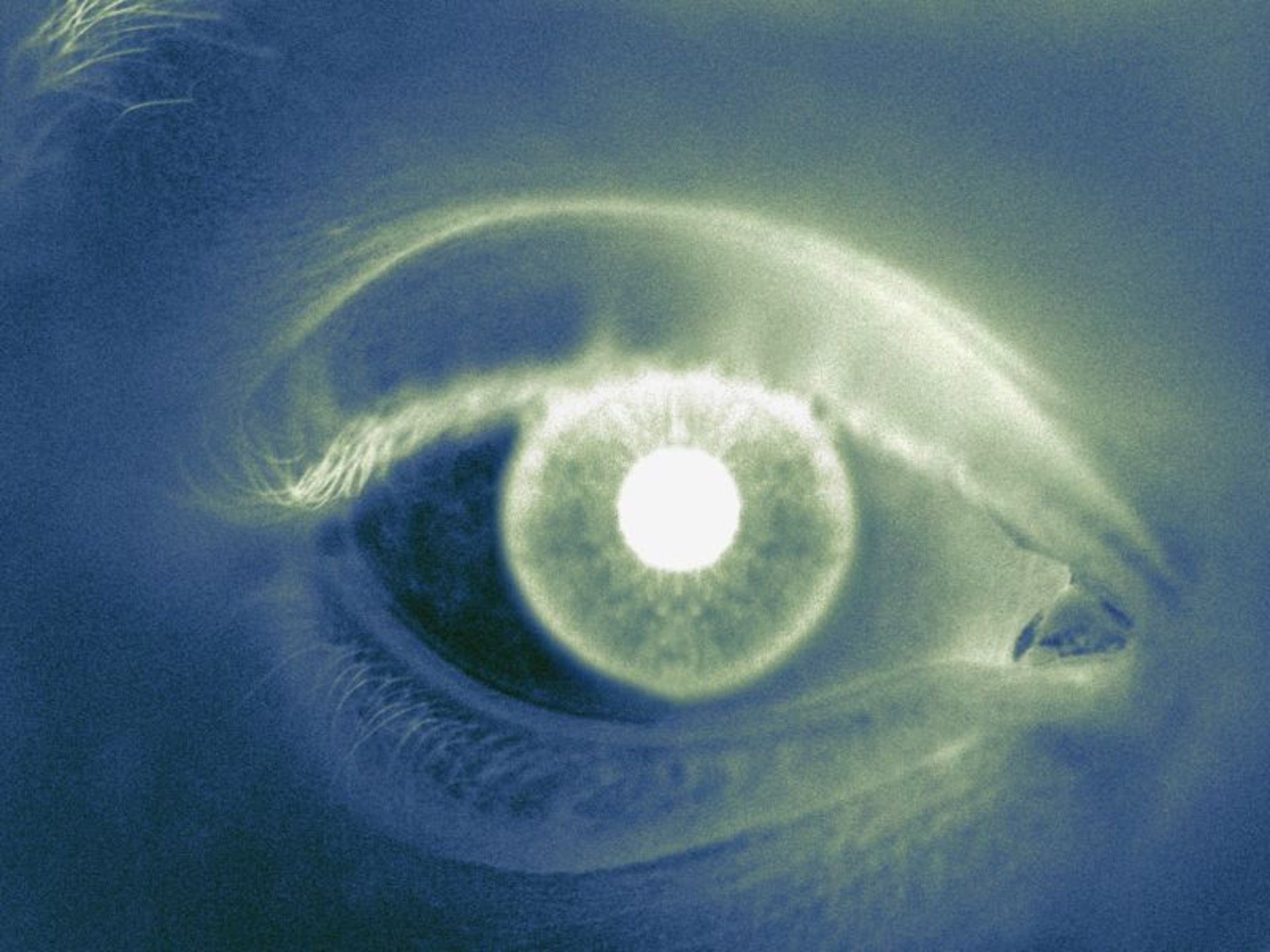
Structural and vascular changes detected on noninvasive retinal imaging tied to performance on tests of cognitive function
WEDNESDAY, Jan. 20, 2021 (HealthDay News) — Abnormalities detected on noninvasive retinal imaging are associated with markers of cognitive dysfunction in older individuals with type 1 diabetes, according to a study published online Dec. 30 in the Journal of Clinical Endocrinology & Metabolism.
Ward Fickweiler, M.D., from the Joslin Diabetes Center in Boston, and colleagues conducted complete cognitive testing, optical coherence tomography (OCT), and OCT angiography (OCTA) on 129 participants with 50 or more years of type 1 diabetes.
The researchers found that decreased vessel density of the superficial retinal capillary plexus and deep retinal capillary plexus was associated with worse delayed memory and dominant hand psychomotor speed. Worse psychomotor speed in both nondominant and dominant hands was associated with thinning of the retinal outer nuclear layer. Delayed memory was associated with outer plexiform layer thickness.
“These findings suggest that noninvasive retinal imaging using OCT and OCTA may assist in estimating the risks for cognitive dysfunction in people with type 1 diabetes,” the authors write.
Abstract/Full Text (subscription or payment may be required)
 Urine Cell-Free RNA Shows Promise as Biomarker for Bladder Cancer
  Age-, Joint-Specific Ultrasound Reference Values Set for Healthy Joints
  Retinal Imaging Can ID Atrial Fibrillation Years Before Diagnosis
  U.S. Adults May Not Understand Their Risk for Vascular Disease
  Lower Cognitive Functioning During 60s Linked to Subsequent Stroke Risk
  Polypharmacy of Prescription Medications Increased for U.S. Older Adults
  Nearly 1 in 10 Older Adults Used Cannabis in the Past Year
  Lower Vertebral Bone Mineral Density Linked to Cognitive Decline



.jpg)